
Cancer Animal Models Shared Resource
The Cancer Animal Models Shared Resource assists investigators in developing, characterizing and analyzing suitable animal models for cancer research.
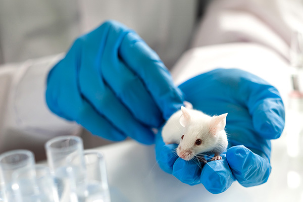
The Cancer Animal Models Shared Resource assists investigators in developing, characterizing and analyzing suitable animal models for cancer research.
The Cancer Animal Models Shared Resource assists investigators in developing and analyzing appropriate animal models, offers cutting-edge imaging modalities for cancer research, and plays an important role in translating research discoveries into potential treatments.
The facility's major emphasis and expertise lie in genetically engineered mouse models, patient-derived tumor xenografts and world-class preclinical imaging modalities. We can customize our services as requested to support development of animal models in a timely and cost-efficient manner.
Our lab facility is located in the Winship Shared Resources Hub on the 3rd floor of The Emory Clinic Building B.
We offer the following services to investigators:

The XPERT® 80 Cabinet X‑ray System delivers high-resolution X-ray imaging across an extra-large field of view within a fully shielded mobile platform. Equipped with a 90 kV source, the system enables rapid imaging of samples of various sizes without compromising resolution. The XPERT 80 is well suited for small animal studies, allowing researchers to track and assess structural changes in bone and detect subtle differences with exceptional clarity. Applications include skeletal imaging and bone mineral density analysis in small animal models.
Please contact us before planning a study with the XPERT 80, as instrument-specific training is required before use.
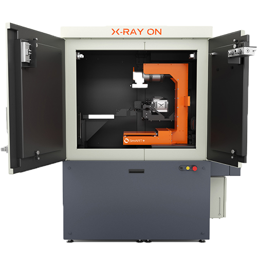
The SmART+ is an advanced platform for image-guided radiation therapy research, enabling precise, targeted irradiation across a wide range of preclinical applications. The system integrates focal irradiation with cone-beam CT, micro-CT (µCT) and bioluminescence imaging to support accurate treatment planning and image-guided radiation delivery. Following image acquisition, the treatment planning system allows researchers to define targets and deliver radiation using fixed or variable collimators with high precision.
We recommend contacting us before planning any study with the SmART+ instrument. For in vivo use of this instrument, please amend the IACUC protocol first.

The IVIS® Spectrum Series is a preclinical optical imaging platform that combines high-throughput imaging with tomographic optical imaging capabilities. The system supports noninvasive, longitudinal monitoring of disease progression, cell trafficking and gene expression in living animals. Highly sensitive bioluminescence imaging, combined with tunable fluorescence imaging and near-infrared (NIR) optimized illumination, enables detection and localization of optical signals from deep tissues, supporting a wide range of in vivo research applications.
The National Cancer Institute requires that publications acknowledge the Winship Cancer Institute Cancer Center Support Grant (CCSG), and it is tracking compliance. If a Winship Cancer Institute CCSG-supported Shared Resource provided data, designed the study, performed analyses, provided results used in your publication, and/or provided any systems or services that were used for the work that resulted in your publication, please include the following statement in the acknowledgment section of your publication(s):
Research reported in this publication was supported in part by the Cancer Animal Models shared resource of Winship Cancer Institute of Emory University and NIH/NCI under award number P30CA138292. The content is solely the responsibility of the authors and does not necessarily represent the official views of the National Institutes of Health.
Winship Cancer Institute of Emory University

Winship Cancer Institute of Emory University