The Cancer Tissue and Pathology Shared Resource facilitates discovery by providing high-quality human cancer specimens, comprehensive histopathologic and immune monitoring services to cancer researchers.
The Cancer Tissue and Pathology Shared Resource is intended and designed to help both individual and team science projects across all major and developing programs within Winship Cancer Institute of Emory University.
The shared resource provides three main services to support basic, clinical and translational research:
Tissue biobanking
Histopathologic tissue
Immune monitoring
Tissue Biobanking Services
The tissue biobank collaborates with all major disease groups at Winship Cancer Institute to collect, store and distribute tumor and normal control tissues for research. Biospecimens are collected under a central institutional review board-approved biobank protocol or under disease-specific or specialized protocols. Specimens are procured as frozen, fresh, fixed, embedded or in other specialized preparations. Tissues and biospecimens are stored and distributed to Winship investigators with institutional review board-approved protocols to support clinical, translational, basic and epidemiologic research. Members of the Department of Pathology provide quality assurance for all specimens.
The laboratory can process high volumes of tissue samples derived from remnant surgical specimens and works in close collaboration with members of the main and developing scientific programs at Winship. The tissue biobank includes an institutional review board-approved Human Tissue Procurement Service, located within Emory University Hospital and Winship Cancer Institute, with affiliated banking sites at Grady Memorial Hospital and Winship Cancer Institute at Emory Midtown.
Histopathologic Tissue Services
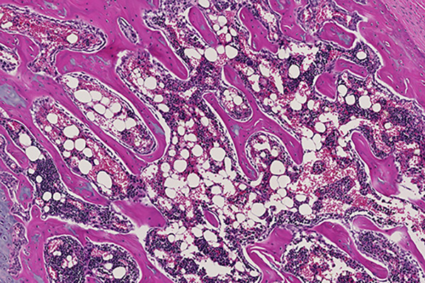
The Cancer Tissue Pathology Lab offers comprehensive histopathologic services ranging from tissue embedding and cutting to multiplex immunohistochemistry. The laboratory can provide nearly all tissue-based needs of investigators.
It is equipped with the following:
Automated tissue processor
Embedding station
Three microtomes and water baths
Cryostat
Automated slide labeler
Automated stainer and coverslipper
Automated single and multiplex immunohistochemical stainer
Manual tissue microarrayer
Semi-automated microarrayer
Our lab facility is located in the Winship Shared Resources Hub on the 3rd floor of The Emory Clinic Building B.
For digital pathology, the lab provides:
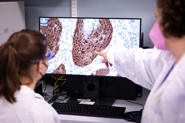
Olympus Nanozoomer whole-slide scanner
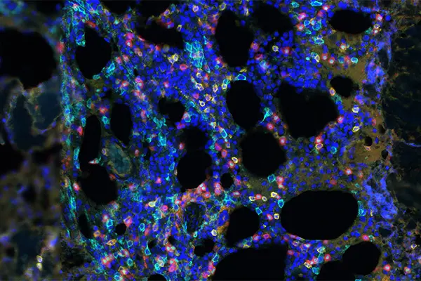
Vectra Polaris multispectral imaging system
Additional equipment includes:
A fume hood for manual histologic staining of cryostat sections or benchtop IHC staining.
A two-headed microscope with digital camera and imaging software for image capturing.
Immune Monitoring Services
Winship members can apply mass cytometry data analysis in applications including:
Cell identification
Detection of clinical biomarkers
Cell phenotyping, phosphorylation-induced signaling and functional readouts of cell-cycle status
Proliferation and apoptosis in human and murine samples
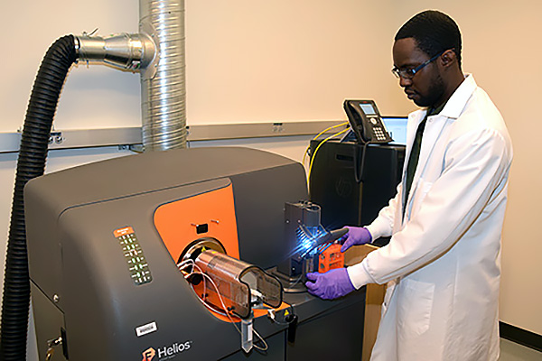
We can measure over 40 available channels simultaneously using metal conjugated antibodies. Users can access our Helios mass cytometer and receive expert counseling on basic science and clinical research applications in the fields of immunology, cancer biology, systems biology and autoimmunity.
In addition, we also offer:
Counseling for panel design, antibody selection and antibody conjugation.
Mass cytometry standardization training for published techniques including antibody titration, data validation and data analysis workflow.
Software support and training for the cloud-based data analysis platform Cytobank.
Be sure to contact us well in advance if you are a first-time user of mass cytometry instrumentation.
Lastly, Winship investigators can access our CTL S6 ImmunoSpot Reader for analysis and training. The reader is a multifunctional, high-throughput imaging instrument for automated ELISPOT and FluoroSpot assay analysis. Its versatile system allows laboratories to switch between microscopic single-cell analysis and macroscopic applications such as viral plaque and bacterial colony counting.
Publication Acknowledgement Policy
The National Cancer Institute requires that publications acknowledge the Winship Cancer Institute Cancer Center Support Grant (CCSG), and it is tracking compliance. If a Winship Cancer Institute CCSG-supported Shared Resource provided data, designed the study, performed analyses, provided results used in your publication, and/or provided any systems or services that were used for the work that resulted in your publication, please include the following statement in the acknowledgment section of your publication(s):
Research reported in this publication was supported in part by the Cancer Tissue and Pathology Shared Resource of Winship Cancer Institute of Emory University and NIH/NCI under award number P30CA138292. The content is solely the responsibility of the authors and does not necessarily represent the official views of the National Institutes of Health.